
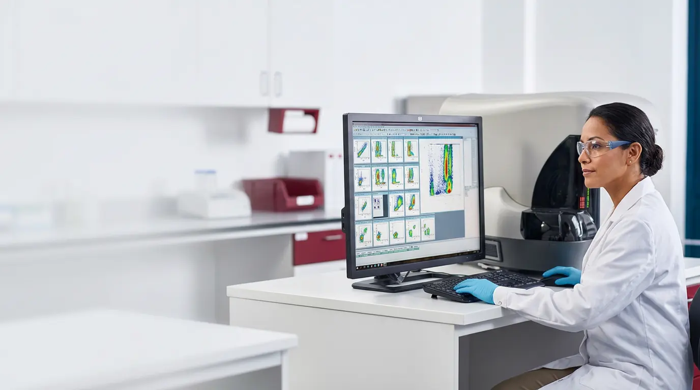
HAIMA · Flow cytometry laboratory
Precise, reliable and timely hematologic diagnosis
Multiparametric flow cytometry immunophenotyping integrated with morphology and cytochemistry. Comprehensive diagnostics in leukemias, lymphomas and cell monitoring.
 Navios EX · 10 colors
0colors
Simultaneous acquisition on Navios EX
0disciplines
Cytometry, morphology and cytochemistry
0years
Institutional experience in cytometry
1–3days
Estimated turnaround by study
Flow cytometry service
HAIMA's flow cytometry laboratory combines multiparametric instrumentation and scientific expertise to deliver reliable results for physicians and hospitals.
From straightforward cell profiling to broad immunophenotyping studies, the panel is defined with the requesting physician around the clinical question.
Foundation of the technique
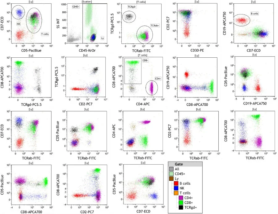
Multiparametric cellular analysis, cell by cell
The analysis combines optical signals and specific markers to identify cell populations and correlate them with morphological and cytochemical findings.
- 01
Staining and lysis
The sample is labeled with specific antibodies and subjected to red-cell lysis until a cell suspension suitable for optical reading is obtained.
- 02
Acquisition cell by cell
Cells travel in an ordered suspension and cross the cytometer's laser beam one at a time.
- 03
Multiparametric reading
Detectors record each cell's response to light, and the resulting parameters are correlated with identifiable cell populations.
Processed samples
Bone marrow · Peripheral blood · Apheresis products · Body fluids · Cell suspensions from tissues with suspected neoplastic infiltration
Clinical catalog
Available immunophenotyping studies
The laboratory confirms the panel, sample type and shipping conditions before collection.
01Leukemia and lymphoma immunophenotyping
Determines lineage and maturation to classify and type hematologic neoplasms.
Typical sample
Bone marrow or peripheral blood, according to the clinical case.
- Initial screening panel: CD45/SSC, CD3, CD19, CD34, CD117, CD56 and HLA-DR.
- B-ALL: B markers, immaturity and maturation stages.
- T-ALL: T markers and cortical stage.
- AML: myeloid, monocytic or megakaryocytic lineage.
- Chronic B lymphoproliferative syndromes: CLL, mantle cell, follicular and hairy cell.
02Lymphocyte subsets (CD4 / CD8 profile)
T-lymphocyte quantification for the patient's immunological monitoring.
Typical sample
Peripheral blood with the anticoagulant indicated by the laboratory.
- Absolute count and percentage of total T lymphocytes (CD3+).
- T helper cells (CD3+CD4+) and cytotoxic T cells (CD3+CD8+).
- CD4/CD8 ratio.
- Common indications: HIV follow-up, immunodeficiencies and transplants.
03CD34+ enumeration (stem cells)
Hematopoietic progenitor count oriented to bone marrow transplant evaluation.
Typical sample
Peripheral blood, bone marrow or apheresis product, according to indication.
- ISHAGE-based enumeration protocol.
- Applicable to peripheral blood, bone marrow and apheresis products.
- Supports collection and transplant timing decisions.
04Measurable residual disease (MRD)
Fine detection of residual abnormal cell populations after treatment.
Typical sample
Bone marrow; collection timing depends on the treatment protocol.
- Applied in B-ALL, AML and multiple myeloma.
- Up to 100 times more sensitive than conventional morphology: detects as few as 1 abnormal cell in 100,000.
- Requires prior diagnostic findings and advance coordination with the laboratory.
05DNA index (ploidy)
Cell ploidy analysis through DNA staining, with prognostic impact in leukemias.
Typical sample
A representative cell population with an appropriate control.
- Determines hyperdiploidy or hypodiploidy.
- Evaluation of cell-cycle distribution.
- Results are integrated with the remaining hematologic findings.
06Platelet antibody detection
Supports the workup of platelet disorders and platelet refractoriness.
Typical sample
Sample and preparation depend on the requested assay.
- Broad antibody panel selected according to the clinical suspicion.
- Direct detection (PAIgG).
- MAIPA: identifies the target platelet glycoprotein.
- Results must be interpreted within the clinical context.
The HAIMA method
Every diagnosis integrates immunophenotype, morphology and cytochemistry—peroxidase and PAS—reviewed together by the specialist and correlated with the patient's clinical data.
Speed without sacrificing quality.
Request and submission
From medical order to result delivery
- 01
Medical order
Order issued by the hematologist or the treating physician.
- 02
Requirements confirmation
The laboratory confirms sample type, tube and transport conditions before collection.
- 03
Collection and transport
HAIMA coordinates local collection or regional sample receipt.
- 04
Result delivery
Estimated turnaround of 1 to 3 days, depending on the type and scope of the study.
Coverage and receipt
Where each sample is received
Analysis is performed at the HAIMA laboratory in Tijuana. Ensenada operates as a receipt point and Mexicali will join the network shortly.
To coordinate collection or shipment of a sample, contact the laboratory before collection.
Tijuana
Operating site
HAIMA Blood Bank · flow cytometry laboratory
Av. Camino Nuevo 2063, Zona Este22000 Tijuana, Baja California
Central laboratory. Local collection at hospitals and hematology practices, and direct sample receipt.
DirectionsEnsenada
Receipt point
Centro Hematológico de Ensenada
Av. Álvaro Obregón 918, Zona Centro22800 Ensenada, B.C.
Consolidates regional samples and coordinates their shipment to the laboratory in Tijuana.
DirectionsMexicali
Coming soon
Regional receipt
Opening soon
Integration into the collection network in preparation; shipment is coordinated with the laboratory in Tijuana.
Recommended preservation
Conditions for a result faithful to the sample
Flow cytometry analyzes living cells. Time and temperature between collection and analysis directly affect cell viability and marker expression.
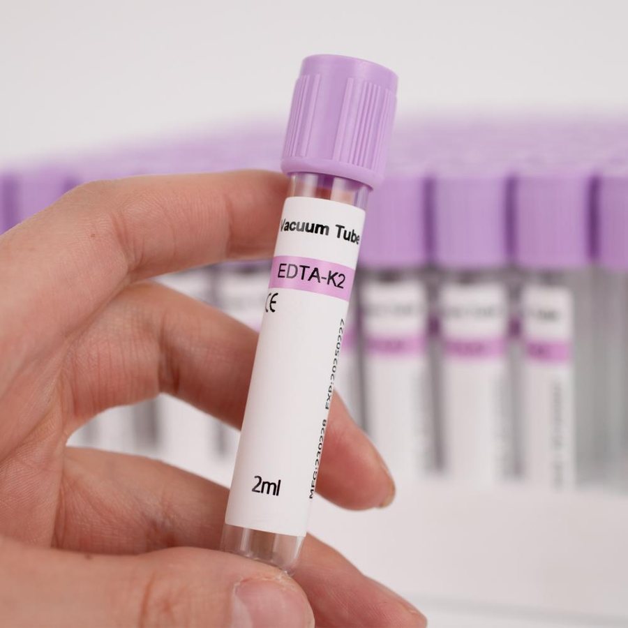
- Tube
- Peripheral blood: lavender-top EDTA tube, gently mixed by inversion after collection.
- Temperature
- Keep at room temperature. Do not refrigerate or freeze unless instructed by the laboratory.
- Time
- Send to the laboratory as soon as possible after collection, preferably the same day. Delays reduce cell viability and may alter the result.
- Transport
- Protect from direct heat, impact and sunlight; include the medical request with the presumptive diagnosis.
- Labeling
- Patient's full name, date and time of collection, and sample type on the tube.
Bone marrow, body fluids, tissues and specialized assays may require different conditions. Confirm the volume and time limits for each study with HAIMA before collection.
Estimated turnaround: 1 to 3 days, depending on the type and scope of the study.
Team and direction
Institutional experience behind every result
Technical team
Mizael Cortes Reyes · Clinical chemist (QFB) · 4 years of experience · Processing and quality control in flow cytometry
Responsible hematologist
Dr. Miguel Guerra · Hematologist, Siglo XXI · Inmunohemato Köln Al. · Member of the Mexican Cytometry Association
Scientific guidance
Dr. Beatriz Pérez Romano · Clinical chemist (QFB) · 30 years of experience
HAIMA track record
At HAIMA: over 25 years of flow cytometry experience.
Reference content for educational/guidance purposes. It does not replace the validated standard operating procedures of each laboratory or current guidelines (WHO/ELN/EuroFlow/ISHAGE).
Direct contact
Study requests and panel confirmation
Indicate the study of interest, presumptive diagnosis and sample type. The laboratory replies with requirements, estimated turnaround and a quote.
Request by study
- Leukemia and lymphoma immunophenotyping
- Lymphocyte subsets (CD4 / CD8 profile)
- CD34+ enumeration (stem cells)
- Measurable residual disease (MRD)
- DNA index (ploidy)
- Platelet antibody detection
Each study is quoted according to sample type, required panel and clinical context.